
Welcome to the Hu Lab for Spatial Systems Immunology @ MD Anderson:
Cleared 250 micron B78 melanoma tumor section imaged for vasculature, myeloid cells, regions of hypoxia and antibody leakage.
Tumors, like other tissues, are composed of a spatially heterogenous arrangement of cells and extracellular elements. Our main goal is to understand how this spatial organization arises, how it interacts with immunotherapies in both helpful and harmful ways, and how we can manipulate it.
We are tissue explorers
Multicellularity demands spatial organization of cells with distinct roles. Tumors are tissues too; can we figure out the guiding principles of their organization and exploit that for therapies? And how can we relate those principles to other tissue contexts such as wound repair? (pictured right)
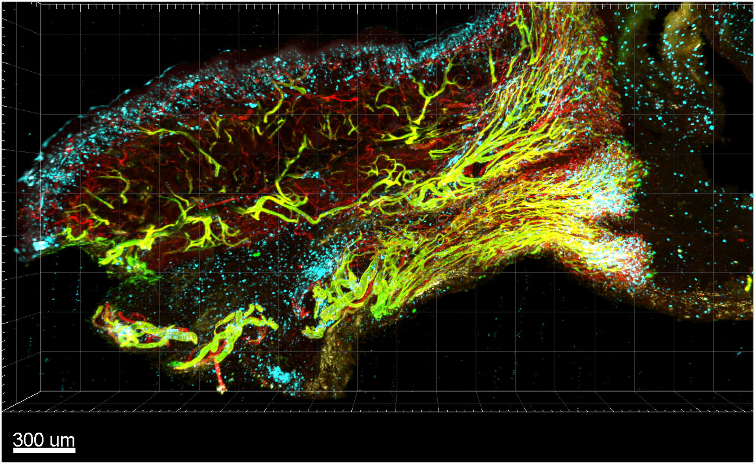
Cleared thick section of mouse wound with CD31+ vasculature in Red, Selectin-P+ vasculature in Green, and MHCII+ cells in Cyan
Animated schematic of the ZipSeq v1 workflow
Unleashing the power of Light
But how do we explore tissues? Light is a remarkable tool not only for imaging, but also for precise spatiotemporal control. We seek new photochemistries for interfacing with biological systems to generate novel methods for dissecting tissue immunology.
Funding Sources:
Thank you to all our generous sources of funding to carry out this work:

